
Track: Orthopedic Radiology
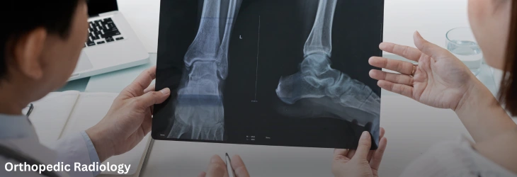
The Orthopedic Radiology session showcases how imaging science continues to evolve as the diagnostic backbone of musculoskeletal medicine in 2027. From high-resolution MRI protocols to weight-bearing CT, attendees will learn how today's imaging tools are catching pathology earlier and with greater precision than ever before.
This session also spotlights the growing role of AI-assisted image interpretation, which is helping radiologists and surgeons alike detect subtle abnormalities faster and more consistently. Case-based discussions will cover imaging strategies for trauma, tumor, and infection workups, along with the pitfalls clinicians should watch for when translating images into treatment plans.
Attendees will leave this session with a sharper understanding of how imaging directly informs surgical decision-making, and how emerging low-dose and point-of-care technologies are making high-quality diagnostics more accessible across different healthcare settings worldwide.
Key Topics and Highlights:
Advanced MRI and CT Techniques: Emerging protocols improving diagnostic clarity for complex musculoskeletal conditions. This session reviews which protocols are gaining traction across leading imaging centers
Weight-Bearing and Functional Imaging: How dynamic imaging is changing surgical planning. Attendees will see case examples where this changed the surgical approach entirely.
AI-Assisted Interpretation: Machine learning tools accelerating and improving image analysis. These tools are increasingly used as a second reader to flag findings for radiologist review.
Trauma & Tumor Imaging Protocols: Best practices for urgent and oncologic imaging workups. Speed and accuracy are both critical in trauma settings, while oncologic imaging demands careful staging protocols.
Infection Workup Imaging: Distinguishing infection from other pathology on modern imaging. Speakers will share imaging patterns and pitfalls specific to periprosthetic infection.
Pitfalls & Pearls: Common misreads and how to avoid them in clinical practice. This session walks through real cases where initial imaging interpretation was later revised.
Scientific Highlights
- Orthopedic Surgery
- AI and Robotics in Orthopedics
- Orthopedic Radiology
- Trauma Reconstruction and Fracture Management
- Complex Arthroplasty and Arthroscopy
- Joint Preservation and Replacement
- Spine Disorders, Deformity and Functional Surgery
- Hand and Upper Extremity Surgery
- Advanced Foot and Ankle Surgery
- Pediatric Orthopedics
- Sports Medicine in Orthopedics
- Orthopedic Oncology
- Orthopedic Infections
- Rheumatology and Arthritis
- Metabolic Bone Diseases and Osteoporosis
- Orthopedic Physiotherapy
- Physical Medicine and Rehabilitation
- Musculoskeletal Disorders: Clinical Trials

Organization Location
1309 Coffeen Avenue, STE 1200, Sheridan, Wyoming 82801, USA
Mail us at
General Enquiry
contact@scientexconferences.com
Call us at
USA : (+1) 312 815 4231
Quick Links
Past Conferences
No past conferences available.
Past Webinar
No past webinars available.